The use of medical imaging in clinical trials has increased in recent years, and computed tomography (CT) is one of the modalities most commonly used. There is a body of literature guiding the measurement of lesions identified on CT scans (eg. RECIST 1.1).
Computed Tomography
The use of medical imaging in clinical trials has increased in recent years, and computed tomography (CT) is one of the modalities most commonly used. There is a body of literature guiding the measurement of lesions identified on CT scans (eg. RECIST 1.1).
However, guidelines regarding the acquisition of CT scans used for the study are usually limited to the study protocol alone and are often written by a someone other than a radiologist. A variety of patient-related and technique-related variables can affect the quality of data acquired and thus ultimately can affect the accuracy of data within the trial. Poor or variable image quality increases the amount of error in each lesion measurement, thus decreasing accuracy of the study. Keeping several basic principles in mind can help to increase data accuracy.
Adherence to the Study Protocol
When an examination is acquired as part of a clinical trial, it is critical to adhere to protocol parameters specified in the study design. While this seems an obvious step in theory, in practice the specified protocol may not always make sense to those who supervise the examinations at participating sites if those individuals were not involved in study design. The use or exclusion of intravenous or enteric contrast, imaging both without and with intravenous contrast or during a specific phase of enhancement may have been selected for a particular reason not immediately apparent to the site and deviations from the protocol may result in data or even patient exclusion. Even if a site has constructive ideas on protocol improvement, a change in the protocol can result in the invalidation of data collected at other sites prior to the change. Thus, protocol revisions are avoided unless absolutely necessary.
Patient-Related Variables
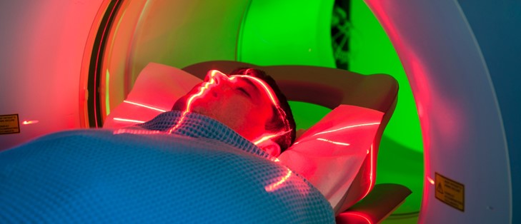
Fortunately, there are relatively few patient-related variables that affect the accuracy or validity of CT scan data used in clinical trials. Perhaps the most important is minimizing patient motion during the scan acquisition. The breath hold remains essential to maintaining scan quality. Despite advances in CT scanner technology that
continue to decrease the time of scan acquisition, a weakened or dyspneic oncology patient may have difficulty holding their breath for even a short period of time. Language barriers may render breathing instructions useless. Motion caused by breathing can result in inaccurate lesion measurements and occasionally results in the appearance of artifact lesions. A diligent technologist who takes the time to coach a patient through a breath hold or finds an interpreter when necessary can help to minimize motion related error.
Technique-Related Variables
In a perfect world, adhering to the parameters specified in the study protocol should result in consistent acquisition technique. However, the process of scan acquisition is often not as simple as it might seem. There is a wide assortment of CT scanners, varying by vendor, detector array, and software version. Most institutions providing data for a single site study have several scanners available. Multicenter trials usually include data acquired from widely different types of CT technology. It is challenging to define technical parameters to achieve comparable image quality with such a broad spectrum of technology. Radiation dose reduction systems designed to optimize the balance between radiation dose and image quality offer some opportunity to achieve consistent image quality among patients of disparate body habitus, but are often difficult to standardize across scanner platforms even within a given institution. Radiologists must work with the technologists at their institution to ensure current modulation programs are being utilized effectively to achieve adequate and consistent lesion conspicuity. Simply having the patient off-center within the gantry can compromise image quality, introducing image noise. Rapid gantry rotation or table speed hinders automated programs from supplying sufficient tube current to achieve acceptable image quality in larger patients. Low pitch protocols can be used to obtain more consistent image quality across a spectrum of patient size. Slice thickness of the reconstructed axial sections affects data accuracy in several ways.
Thinner sections (typically 3 mm or less) are more likely to allow consistent measurement of small lesions (eg. lymph nodes) as slice selection variability is less likely to result in under-measurement. Thicker sections (5 mm), however, usually provide better lesion conspicuity for liver metastases and other potentially low-contrast lesions. These factors should be considered when designing the study and specified in the protocol. If, however, it is not specified in the study protocol, sites are strongly encouraged to adhere to a consistent reconstructed slice thickness for all examinations in a given study. Timing of scan initiation following or during a contrast bolus can be a critical factor to lesion conspicuity. As with tube current, it can be challenging to achieve consistent bolus timing across scanner platforms. For a given patient scanned each time on the same scanner, consistency can usually be achieved by adhering to the same bolus timing protocol used for the last scan on that patient, either a standardized time delay or bolus tracking software. However, if a given patient may be examined on one of several scanners over the course of the study, it is important that the radiologists and technologists have worked together on bolus timing for each scanner so that an arterial, portal venous or other standardized degree of enhancement is achieved consistently.
Summary
Some automated data measurement programs are available to provide 2-D and 3-D assessment of lung nodules as well as organ, lesion and lymph node size. As these tools become more refined, they are likely to play a greater role in data collection in clinical trials. To date, most if not all of these programs utilize some form of “nearest neighbor” algorithm, using similarities and differences between adjacent pixels or voxels to define a lesion. Effective use of such programs still requires attention to the previously described variables that affect image quality, in fact image noise can have significant impact on lesion definition. In addition, accuracy will also require isotropic or at least near-isotropic data to avoid longitudinal inaccuracy in volumetric measurements.
Volume 4, Issue 7: Guidance For Sponsors: Computed Tomography Data Acquisition in Clinical Trials
Originally written by legacy Intrinsic Imaging Medical Director
Contact WCG Imaging to discuss your trial’s imaging needs
We have the team, therapeutic expertise, technology, and ISO-certified quality management systems to provide imaging core lab services to our clients worldwide. Complete the form to get started.